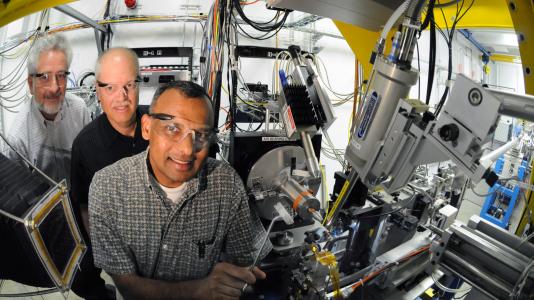
But that may be changing now that researchers have mapped one virus’s atomic structure using the Advanced Photon Source (APS) at the U.S. Department of Energy’s Argonne National Laboratory.
Glen Nemerow and Vijay Reddy have been studying the human adenovirus—responsible for 10 percent of colds in addition to other, more harmful infections—for more than a dozen years.
Nemerow, a professor in the Department of Immunology and Microbial Science at The Scripps Research Institute in La Jolla, Calif., and Reddy, an associate professor in Scripp’s Department of Molecular Biology, paired up in the late 1990s.
Together they mapped the virus using X-ray crystallography, a technique in which X-rays are beamed at a virus crystal, resulting in a diffraction pattern that helps scientists understand the virus’ shape.
Their findings were published in the journal Science on August 27, 2010.
“The more we know about the virus’ structural features, the better we understand how it functions,” Nemerow said. “This will help us learn more about how the virus infects host cells so that we can develop effective anti-virals.”
The major protein component of adenovirus, called hexon, was crystallized—meaning that the proteins were arranged in a way that they could be studied by X-ray diffraction—in 1968. But it wasn’t until 2000 that the refined structure of the hexon could be determined.
By contrast, Nemerow and Reddy determined the structure of the entire virus. This has allowed them to learn how that major protein or hexon was incorporated into the virus and how it interacts with other proteins.
The Scripps scientists said their discovery would not be possible without the use of Argonne’s APS.
“The use of this particular beam line was critical,” Reddy said. “You need to have a high resolution to be able to visualize the virus in greater detail. We could only obtain that at Argonne.”
Reddy said he and Nemerow tried a number of other beam lines before they settled on what is known as the General Medicine and Cancer Institutes Collaborative Access Team (GM/CA-CAT) at the APS.
“The beam line is quite intense compared to others,” Reddy said. “It’s more laser-like.”
Reddy said he and Nemerow value not only the strength of the X-ray but the diligence with which the line is improved and maintained.
“The features of the beam line are absolutely essential,” Nemerow said. “We need a very high energy X-ray beam focused on a very small area to provide the highest resolution data.”
But even before they could get to the APS, the two had to solve nearly insurmountable obstacles.
“Perhaps the most critical step in this process is growing the crystals of the virus,” Reddy said. “That is very important. Not all crystals are able to diffract to a high resolution, and that is a tremendous obstacle in structural biology. It took us five years to cultivate the crystals we needed.”
The pair started coming to Argonne in 2005 and stayed for several days at a time to collect their data.
Robert Fischetti, a senior scientist in the biosciences division at Argonne who has worked with Nemerow and Reddy for years, said that GM/CA-CAT is constantly making improvements to the beam line.
“We spent years perfecting the uniformity of the beam just as they perfected the crystals,” he said.