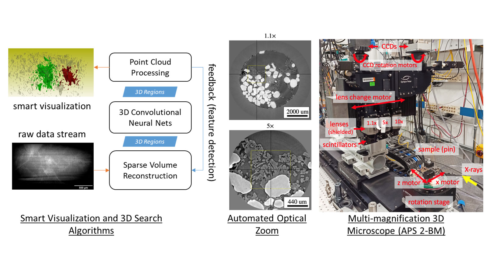
We have developed a software-powered, three-dimensional (3D) X-ray microscope with AI-based computer vision algorithms that is as easy to use as a smart phone.
Our software integrates (1) tomographic (CT) reconstruction; (2) neural networks for segmentation or feature detection; and (3) computational geometry algorithms for tasks such as real-time visualization, automated 3D optical-zoom to regions of interest, and smart capture of data during time-resolved 3D imaging experiments.
We demonstrated prototype solutions through 2021 with select Advanced photon Source (APS) users at the 2-BM and 7-BM beamlines. The software is now available in plug-n-play for all beamlines using the EPICS interface for instrument control.
More recently, we also demonstrated a similar solution, “ROI-Finder,” for automatically recommending scan locations for X-ray Fluorescence Microscopy at the 9-ID bio-nanoprobe at APS. “AI-steer” is a targeted software development effort supported by Argonne’s computational and beamline scientists. “Tomo2Mesh” is a code for real-time reconstruction, segmentation, visualization of raw tomographic data for X-ray micro-CT instruments at APS, such as 2-BM, 7-BM and 1-ID. The project is employing datasets and access to beamtime through contributions and collaborations from APS users.