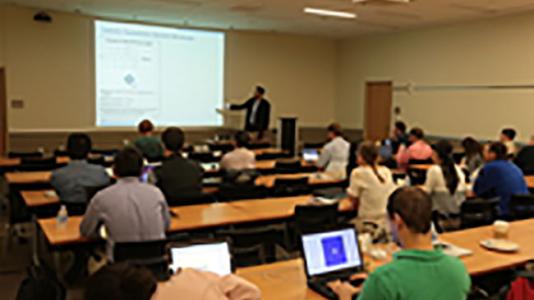
The workshop was entitled “Tomography and Ptychography,” and it brought together researchers in experimental imaging, image and data analysis, and visualization. The workshop acted as a catalyst for new collaborations across Argonne and with external researchers, highlighted Argonne’s imaging expertise and capabilities, and introduced non-experts in imaging to the ways in which these techniques could be used.
The workshop included 23 invited talks and roughly 20 posters. Nine of the speakers were from outside Argonne. The Argonne speakers included representatives from the laboratory’s Advanced Photon Source, Materials Science Division, Mathematics and Computer Science Division, and Argonne Leadership Computing Facility. The workshop attracted 92 attendees.
The workshop focused on the presentation of tomography and ptychography applications based on different imaging technologies including X-ray microscopy, electron microscopy, and atom probe tomography. The researchers discussed applications that included mechanical alloys, geoscience, spatially-confined surface plasmons on metallic nanoparticles, magnetic nanostructures, targeted drug delivery for cancer cells, magnetotactic bacteria, gallium-nitride light-emitting diodes, and the strain distribution inside zinc oxide nanopillars.
The workshop speakers also discussed some of the challenges associated with processing and visualizing the complex data sets that are acquired using these techniques. For example, samples may be sensitive to the electrons or X-rays used to image them, leading to a need to calculate tomography or ptychography reconstructions from less than perfect initial data sets. Examples of ways to solve this include the use of compressed sensing for tomography, or using a constantly moving X-ray beam to scan over a sample so that the exposure time at each point is reduced.
Further challenges are associated with transferring data from the microscope where it is collected to computers used for data analysis and processing, which can be time-consuming for the very large data sets that are typically obtained. The scientists also examined how to combine data from different imaging modalities in order to get a better reconstruction of the sample, as well issues associated with visualizing 3D data sets and in extracting quantitative information from the reconstructions.