Scientific Achievement
A penalized maximum-likelihood estimation is proposed to perform hyperspectral (spatio-spectral) image reconstruction for X-ray fluorescence tomography. The resulting element distribution estimates with the proposed approach show significantly better reconstruction quality than the conventional analytical inversion approaches, and allows for a high data compression factor which can reduce data acquisition times remarkably. In particular, this technique provides the capability to tomographically reconstruct full energy dispersive spectra without compromising reconstruction artifacts that impact the interpretation of results.
Significance and Impact
Hard X-ray fluorescence (XRF) tomography has grown into a powerful non-destructive technique for probing trace metal distributions in samples without the need for physical sectioning that may disrupt trace element distributions and sample structure. The technique is becoming increasingly important in a growing number of research fields. A key advantage of XRF imaging is its high sensitivity and specificity for transition metals such as iron, copper, zinc, or other essential trace elements, its high resolution that allows imaging at scales from few microns down to tens of nanometers. The results published to date have largely used conventional filtered back-projection reconstruction methods. While these have yielded very good results, the ultimate quality and precision of the inversion is dependent on both theoretical and experimental factors, such as dealing with finite sampling of data and measurement noise. In this work a hyperspectral inversion approach is presented to recover the full energy dispersive spectra. This approach avoids the conventional reconstruction artifacts that impact interpretation of results. Besides it allows for a high data compression factor which can significantly reduce data acquisition times, and improve beamtime efficiency.
High-Level Research Details
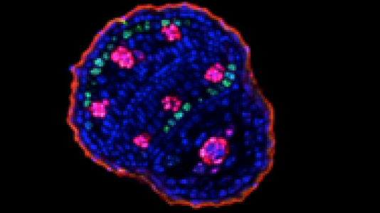
The approach minimizes a Poisson-based negative log-likelihood of the observed photon counts, and uses a penalty term that has the effect of encouraging local continuity of model parameter estimates in both spatial and spectral dimensions simultaneously. The performance of the reconstruction method is demonstrated with experimental data acquired from a seed of arabidopsis thaliana collected at the 13-ID-E microprobe beamline at the Advanced Photon Source.
Additional Research Details
The proposed reconstruction algorithm is implemented on a high-performance data-intensive computing middleware and performed image reconstruction at Argonne Leadership Computing Facility. We ran our experiments on Mira, a 10-petaflops IBM Blue Gene/Q system. We used up to 896 nodes where each node consists of 16 physical cores (14336 cores in total) and 16 GiB memory. The running time of the total hyperspectral reconstruction takes 6.77 seconds for 1 sPML iterations.
Research Team
Doga Gursoy (Argonne X-ray Science Division), Tekin Bicer (Argonne Mathematics and Computer Science Division), Antonio Lanzirotti (Center for Advanced Radiation Sources, University of Chicago), Matthew G. Newville (Center for Advanced Radiation Sources, University of Chicago), Francesco De Carlo (Argonne X-ray Science Division)
Sponsors
U.S. Department of Energy: Office of Science, Geosciences; National Science Foundation: Earth Sciences.
References
- D. Gursoy, T. Bicer, A. Lanzirotti, M. G. Newville and F. De Carlo, “Hyperspectral image reconstruction for x-ray fluorescence tomography,” Optics Express, vol. 23 (2015).
Acknowledgments
We thank Amanda Socha and Tracy Punshon (Dartmouth College) for sharing the arabidopsis thaliana data used in this paper. We also thank Stefan Vogt, Chris Jacobsen, Eugene Lavely, and Yi-San Lai for fruitful discussions and helpful comments during the course of the work.