Scientific Achievement
We describe a new combined development in the field of imaging science and microscopy, with a high spatial resolution of sub-30 nm for x-ray imaging of a frozen hydrated cell. The work combines ptychography (a form of coherent diffraction imaging) to view cellular structure in whole cells too thick for electron microscopy and at higher resolution for unlabeled structure than can be achieved in visible light microscopy, and x-ray fluorescence used to view elemental content with thousandfold higher sensitivity than in electron microprobes. By working with frozen hydrated cells at liquid nitrogen temperature, we obtain high fidelity structural and chemical preservation.
Significance and Impact
The combination of cryogenic ptychography and x-ray fluorescence microscopy provides the capability of obtaining simultaneous views of ultrastructure and elemental compositions of specimens at high resolution. This combined approach will have significant impact in studies of the intracellular localization of nanocomposites with attached therapeutic or diagnostic agents, help elucidate the roles of trace metals in cell development, and further the study of diseases where trace metal misregulation is suspected (including neurodegenerative diseases).
High-Level Research Details
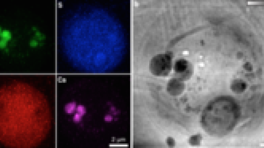
X-ray fluorescence microscopy (XFM) offers high sensitivity for quantitative mapping of elements in biological samples. However, a majority of biological structures are not easily visualized in XFM since XFM is relatively blind to the light elements (such as H, C, N, O) which are the main constituents of biological materials. Ptychography, a more recent method of coherent diffraction imaging (CDI) that delivers quantitative phase contrast and high spatial resolution, provides a good solution to visualize biological ultrastructure. While the spatial resolution of ptychography can in theory reach the wavelength limit, radiation damage to biological samples affects the obtainable resolution. A good solution towards this problem is to work with frozen-hydrated biological specimens under cryogenic conditions, since cryogenic conditions are known to provide excellent structural preservation and radiation damage resistance and to fully preserve chemical integrity of the specimen.
We showed the use of two imaging techniques at once on algae cells that were quickly frozen from the living state, and obtained complementary information from the sample: structural information at sub-30 nm resolution by ptychography, and x-ray fluorescence measurements of chemical element distributions at sub-90 nm resolution. The ptychography reveals in amazing detail the organelles and membranes, which do not fluoresce. By setting the elemental signals in the same context with high-resolution biological structures given by ptychography, the analysis of trace elements can be improved.
Research Team
Junjing Deng and Qiaoling Jin (Northwestern University), Chris Jacobsen (Northwestern University and Argonne X-ray Science Division), David Vine, Si Chen and Stefan Vogt (Argonne X-ray Science Division), Youssef Nashed, Tom Peterka and Rob Ross (Argonne Mathematics and Computing Science Division), Nicholas Phillips (LaTrobe University, Australia)
Sponsors
U.S. Department of Energy: Office of Science. NIH: National Institute of General Medical Sciences. NIH: National Center for Research Resources.
References
- J. Deng, D. J. Vine, S. Chen, Y. S.G. Nashed, Q. Jin, N. W. Phillips, T. Peterka, R. Ross, S. Vogt, and C. Jacobsen, “Simultaneous cryo x-ray ptychographic and fluorescence microscopy of green algae,” PNAS, vol. 112 (2015).
Acknowledgments
We thank R. Mak and M. Guizar-Sicairos for valuable discussions, and K. Brister, C. Roehrig, J. VonOsinkski, and M. Bolbat for help during the experiments. We thank NIH National Institute of General Medical Sciences for support of this work under Grant 1R01GM104530. The Bionanoprobe is funded by NIH/National Center for Research Resources High End Instrumentation Grant 1S10RR029272-01 as part of the American Recovery and Reinvestment Act. Use of the Advanced Photon Source, an Office of Science User Facility operated for the US Department of Energy (DOE) Office of Science by Argonne National Laboratory, was supported by the US DOE under Contract DE-AC02-06CH11357.