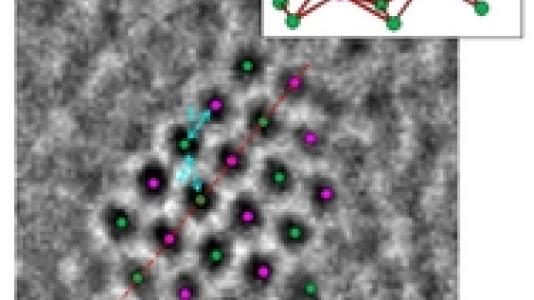
Aberration-corrected (AC) STEM, AC-TEM and in situ X-ray absorption fine structure spectroscopy (XAFS) were used by users from UOP-Honeywell, working with researchers in the CNM Electron Microscopy Center and APS, to characterize Pt clusters on a 0.35 wt % Pt on g-alumina support after reduction in hydrogen at 700°C. STEM high-angle annular dark field imaging shows that cluster formation takes place at temperatures up to approximately 350°C, and this is followed by gradual growth in cluster size for heat treatments in hydrogen up to 700°C. STEM data show that after 700°C reduction the Pt clusters are present in a narrow size distribution centered at 0.88 nm, and using a method involving a redistribution of the Pt atoms using a high electron dosage in the STEM, it is shown that the clusters are present in two-dimensional morphology. This conclusion is verified using intensity line scans. The in situ extended X-ray absorption fine structure data are in good agreement with these observations. High-resolution AC–TEM, which uses a broad coherent electron beam, and can thus offer advantages relative to STEM for structure determination of fine clusters, supported by image simulations of through-focus series, were used to analyze the structures of Pt particles. The structures determined using AC–TEM are consistent with STEM and EXAFS data in having a flat two-dimensional morphology. Comparison of AC–STEM and AC TEM data for the same 700°C reduced sample suggests that parallel-beam TEM mode of imaging may be advantageous because of the less pronounced beam-induced structural rearrangements that occur when imaging with a fine STEM probe.
A comparison of advanced microscopy methods for imaging platinum (Pt) catalyst nanoparticles suggests the aberration-corrected transmission electron microscopy (AC-TEM) mode may be advantageous because of the less pronounced beam-induced structural changes that occur when imaging with a fine scanning TEM (STEM) probe. The possibility of directly determining structures of individual Pt nanoclusters is of direct relevance to understanding the changes in cluster evolution with temperature, which may be relevant for performance in key industrial catalytic processes. Aberration-corrected STEM performed at UOP LLC, AC-TEM performed at CNM, and in situ X-ray absorption fine structure spectroscopy (XAFS) carried out at APS were used to characterize Pt clusters on a 0.35 wt % Pt on gamma-alumina support after reduction in hydrogen at 700°C. The apparent advantage of AC-TEM is due to a broad coherent electron beam.
“Aberration-Corrected Transmission Electron Microscopy and In Situ XAFS Structural Characterization of Pt/g-Al2O3 Nanoparticles ”, W. Sinkler et al., ChemCatChem, 7, 3779 (2015)