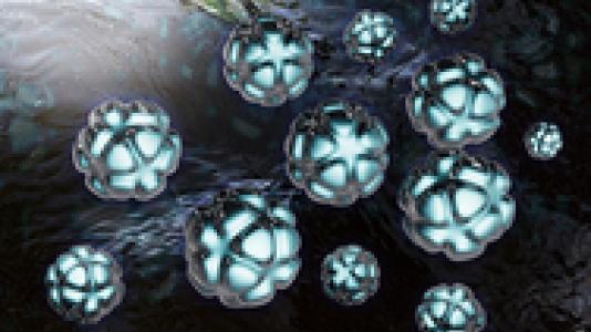
By combining magnetic nanoparticles with a common chemotherapy drug, users from Argonne’s Materials Science Division and the University of Chicago, working with the Center for Nanoscale Materials Nanobio Interfaces and X-ray Microscopy Groups, created a way to deliver anti-cancer drugs directly into the nucleus of cancer cells. The CNM hard X-ray nanoprobe beamline at the APS visualized the platinum distribution using X-ray fluorescence microscopy pinpointed the Pt distribution at the subcellular level at less than 100 nm resolution.
The micelles contain magnetic nanoparticles of iron oxide and cisplatin and can be customized to target specific cancer cells. To carry cisplatin into the cell nucleus, an applied magnetic field causes the encapsulated iron oxide nanoparticles to heat and collapse the polymer micelle, releasing cisplatin. The micelles were created as well as imaged at CNM using a laser scanning confocal fluorescence microscope Zeiss LSM-500 META and a field-emission scannin§g electron microscope JEOL JSM-7500F. The CNM/APS hard x-ray nanoprobe beamline visualized the platinum distribution using x-ray fluorescence microscopy with fine detail at sub-100 nm resolution, pinpointing the Pt distribution at the subcellular level.
This research could potentially increase the amount of cisplatin in cancer cells significantly, making it that much more effective a chemotherapeutic agent.
E.A. Vitol et al., Advanced Materials Interfaces, 1400182 (2014). Online