Express Licensing
The method allows more rapid, yet still non-invasive, detection and is expected to enhance the treatment experience for breast cancer patients.
Because cancer cells grow more quickly than healthy cells, they are typically a few degrees higher in temperature. This attribute makes it possible to use thermal imaging to detect them. For this reason, passive thermal imaging is helpful in detecting breast cancer. In active thermal imaging, heat or cold is applied to an object and an infrared camera is used to observe the resulting temperature change.
Thermal imaging can be used to analyze even multilayered materials— making the methodology even more useful than conventional processes like X-rays, CT and ultrasonic scanning for such applications.
A team of researchers from Rush University Medical Center and Argonne National Laboratory is using a 3-D technique to detect early skin changes due to radiation treatment in breast cancer patients. Use of the technique could facilitate earlier delivery of treatment to prevent radiation skin damage in these patients. They can develop a skin reaction that, in severe cases, causes discomfort and can disrupt therapy. However, if detected early, skin reactions are preventable or treatable.
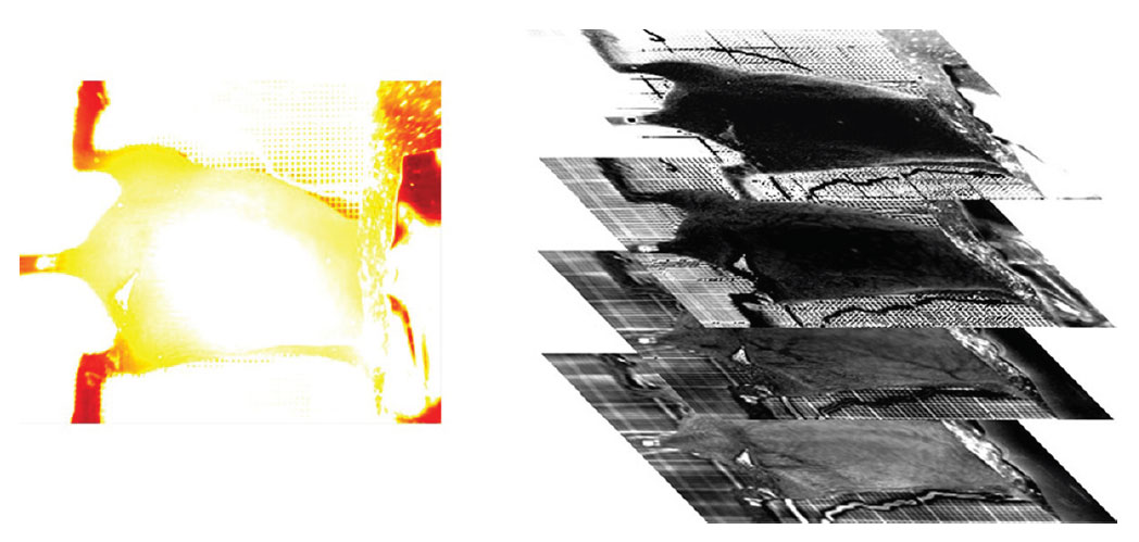
Clinical tests used 3DTT to measure the skin’s thermal effusivity—a tissue property that quantifies its ability to exchange heat with its surroundings. In the test, a flash of filtered light heats the skin while an infrared camera captures a time series of images that display skin temperatures by color. Using an algorithm to calculate temperature changes and determine the thermal effusivity at different skin depths, the researchers discovered the effusivity values of damaged skin tissue differ from that of healthy skin.
Preliminary data show that marked reductions in the effusivity levels of irradiated skin occur well in advance of development of high-grade skin reactions. Soon, the team hopes to apply the 3DTT technique in breast cancer patients. Also underway is the development of an even more sophisticated algorithm to improve resolution at subsurface depths.
Three-dimensional thermal tomography offers significant benefits over existing technologies:
The 3DTT technique has wide application in medicine as well as in other industries where in situ inspection is required, such as the imaging of engine components or the space-shuttle thermal protection system.
This technology is ready for prototype development.