Argonne maintains a wide-ranging science and technology portfolio that seeks to address complex challenges in interdisciplinary and innovative ways. Below is a list of all articles, highlights, profiles, projects, and organizations related specifically to biology.
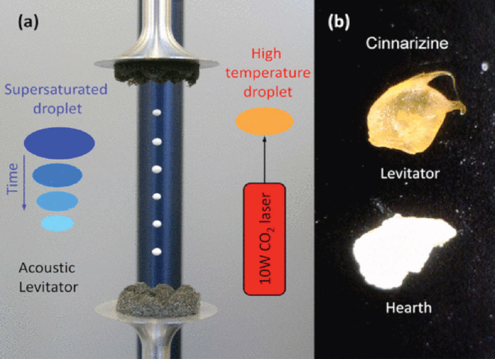
Making fast-acting pharmaceuticals is a goal of almost every drug company. The route of delivering pharmaceuticals in the form of amorphous solids has long been recognized as a possible way to improve dissolution rates and to increase both solubility and bioavailability. Development in this direction is becoming increasingly important due to the emergence of many new drugs that are virtually insoluble in their crystalline form. Researchers at Argonne National Laboratory have developed the technique of acoustic levitation to prepare amorphous solids and molecular gels that can be easily applied to the pharmaceutical manufacturing process. This technique would improve the solubility and bioavailability of several drugs.
The acoustic levitation techniques developed at Argonne keeps the drug solution from making contact with any surface whatsoever during the solvent evaporation process. This containerless process was developed and tested on several over-the-counter and prescription pharmaceuticals. Several of the pharmaceuticals amorphized using the Argonne process remained completely amorphous for four months or longer. Please review the publication for additional details.
A containerless process:
Pharmaceutical industry
Proof of principle
Express Licensing
The method allows more rapid, yet still non-invasive, detection and is expected to enhance the treatment experience for breast cancer patients.
Because cancer cells grow more quickly than healthy cells, they are typically a few degrees higher in temperature. This attribute makes it possible to use thermal imaging to detect them. For this reason, passive thermal imaging is helpful in detecting breast cancer. In active thermal imaging, heat or cold is applied to an object and an infrared camera is used to observe the resulting temperature change.
Thermal imaging can be used to analyze even multilayered materials— making the methodology even more useful than conventional processes like X-rays, CT and ultrasonic scanning for such applications.
A team of researchers from Rush University Medical Center and Argonne National Laboratory is using a 3-D technique to detect early skin changes due to radiation treatment in breast cancer patients. Use of the technique could facilitate earlier delivery of treatment to prevent radiation skin damage in these patients. They can develop a skin reaction that, in severe cases, causes discomfort and can disrupt therapy. However, if detected early, skin reactions are preventable or treatable.
Clinical tests used 3DTT to measure the skin’s thermal effusivity—a tissue property that quantifies its ability to exchange heat with its surroundings. In the test, a flash of filtered light heats the skin while an infrared camera captures a time series of images that display skin temperatures by color. Using an algorithm to calculate temperature changes and determine the thermal effusivity at different skin depths, the researchers discovered the effusivity values of damaged skin tissue differ from that of healthy skin.
Preliminary data show that marked reductions in the effusivity levels of irradiated skin occur well in advance of development of high-grade skin reactions. Soon, the team hopes to apply the 3DTT technique in breast cancer patients. Also underway is the development of an even more sophisticated algorithm to improve resolution at subsurface depths.
Three-dimensional thermal tomography offers significant benefits over existing technologies:
The 3DTT technique has wide application in medicine as well as in other industries where in situ inspection is required, such as the imaging of engine components or the space-shuttle thermal protection system.
This technology is ready for prototype development.
Manipulating electron beam cancer therapy so it can be used treat internal cancers and tumors has the potential to revolutionize oncology. This ground-breaking innovation can provide a successful and cost-effective means of treating cancer in previously inoperable or radiation-sensitive areas of the body.
By delivering large irradiation doses in a short time, electron beams have proven to be very effective in cancer treatment. But the electron is also strongly absorbed by tissue, limiting this treatment to surface cancers and procedures that require large surgical incisions to expose the body core.
Researchers at Argonne National Laboratory, led by John Noonan, have discovered a way to turn the negative attributes of electron beam cancer therapy into advantages. If the electron beam can be transported to the internal cancer without exposure to tissue, the beam can be absorbed by the tumor only. With this approach, healthy tissue is not exposed to radiation.
An electron source has been designed to have very low beam emittance. The beam is sub-millimeter in diameter and stays small over meters of transport in free space. It will allow for an articulated, hard-walled laparoscopic tube to be inserted through a small incision and positioned directly at the tumor. The beam can vary energy from 1 million electron volts (MeV) to 10 MeV, permitting it to cover a tumor size of about 0.5 cm to 5 cm, respectively.
Initially, electron beam treatment can be used on X-ray radiation resistant tumors. The electrons destroy cancerous cells by direct damage to the DNA, and not by electron displacement in molecules as with X-rays. Ultimately, the electron beam therapy would be a competitor to all X-ray treatments.
The damage volume of the electron irradiation can be controlled very closely by changing the electron beam energy. This precise exposure provides several new cancer therapies or treatments in previously inoperable or radiation-sensitive locations, such as the spine, nerves, optic nerve, and organs. Electron beam treatment of brain tumors is another new opportunity. In this case, the laparoscopic tube provides an advantage. After the irradiation, the tube can be used to evacuate the mass of dead tissue, which can become destructive to healthy brain cells.
Enormous doses can be delivered to the tumor without worrying about total body dose exposures, as is required for X-rays. The electron beam can be tailored to irradiate a very precise volume, so an oncologist can direct the irradiation at the tumor and whatever adjacent tissue they feel necessary. Another major advantage over X-rays is the amount of treatment time required. X-ray treatments can go on for months, while electron beams may potentially only require one session, providing a significant improvement in patient care.
The electron beam system is compact so it could fit in an operating room—probably even under the operating table. Except for the electron source, the system uses conventional accelerator technology. The production cost of the unit should be much less than that of existing radiation therapy systems.
Prototype
Noonan J., and Lewellen J.W., 2005, “Field-emission cathode gating for RF electron guns,” Physical Review, Special Topics - Accelerators and Beams 8: 033502.
